At the Seedy Saturday on the weekend where I was selling mason bees, I met Dr. Elaine C. Humphrey, Fellow of Microscopy Society of America, and a Past President Microscopical Society of Canada. She works at the Advanced Microscopy Facility.
Bob Wright Science Centre A015, University of Victoria.
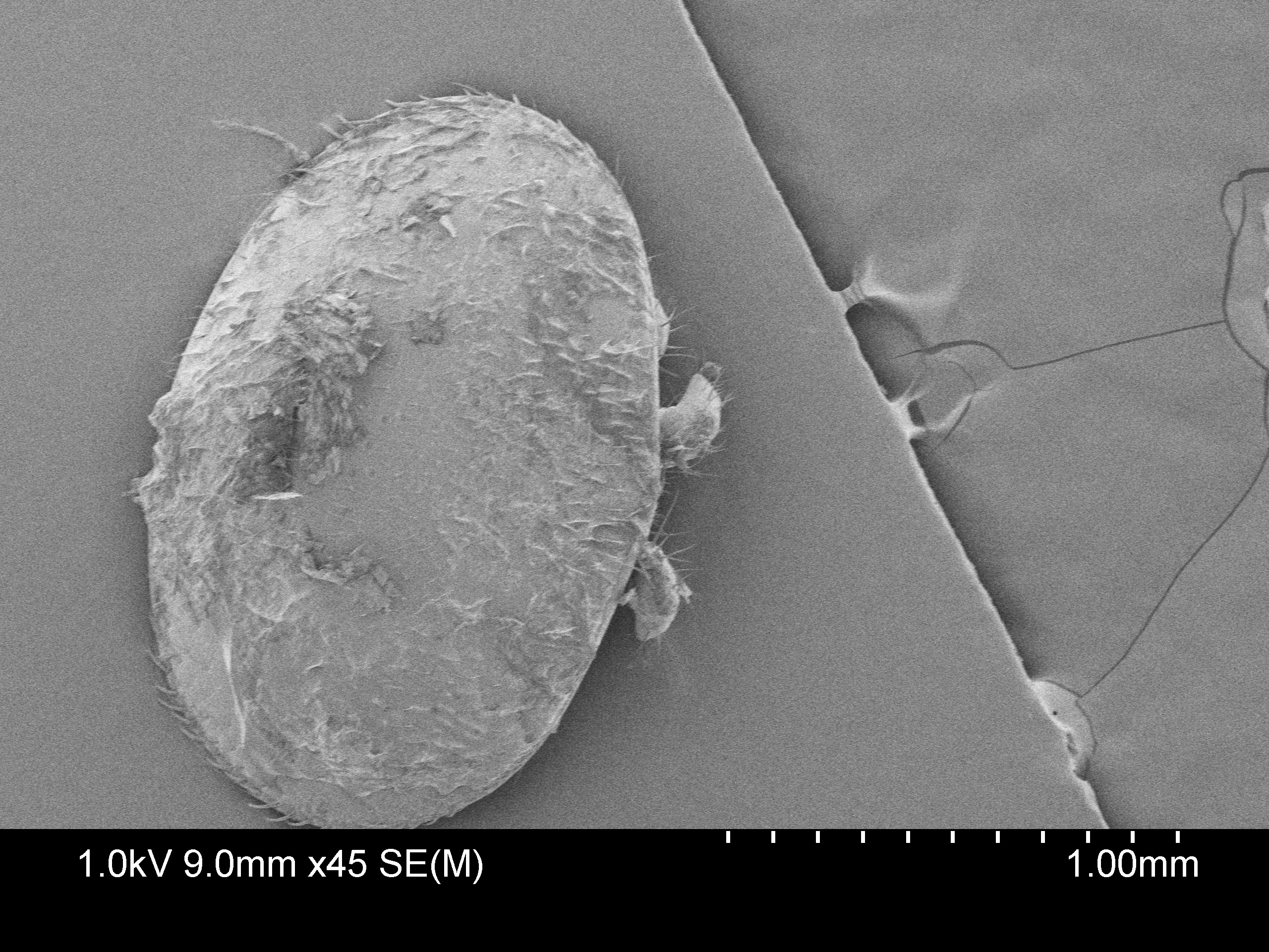
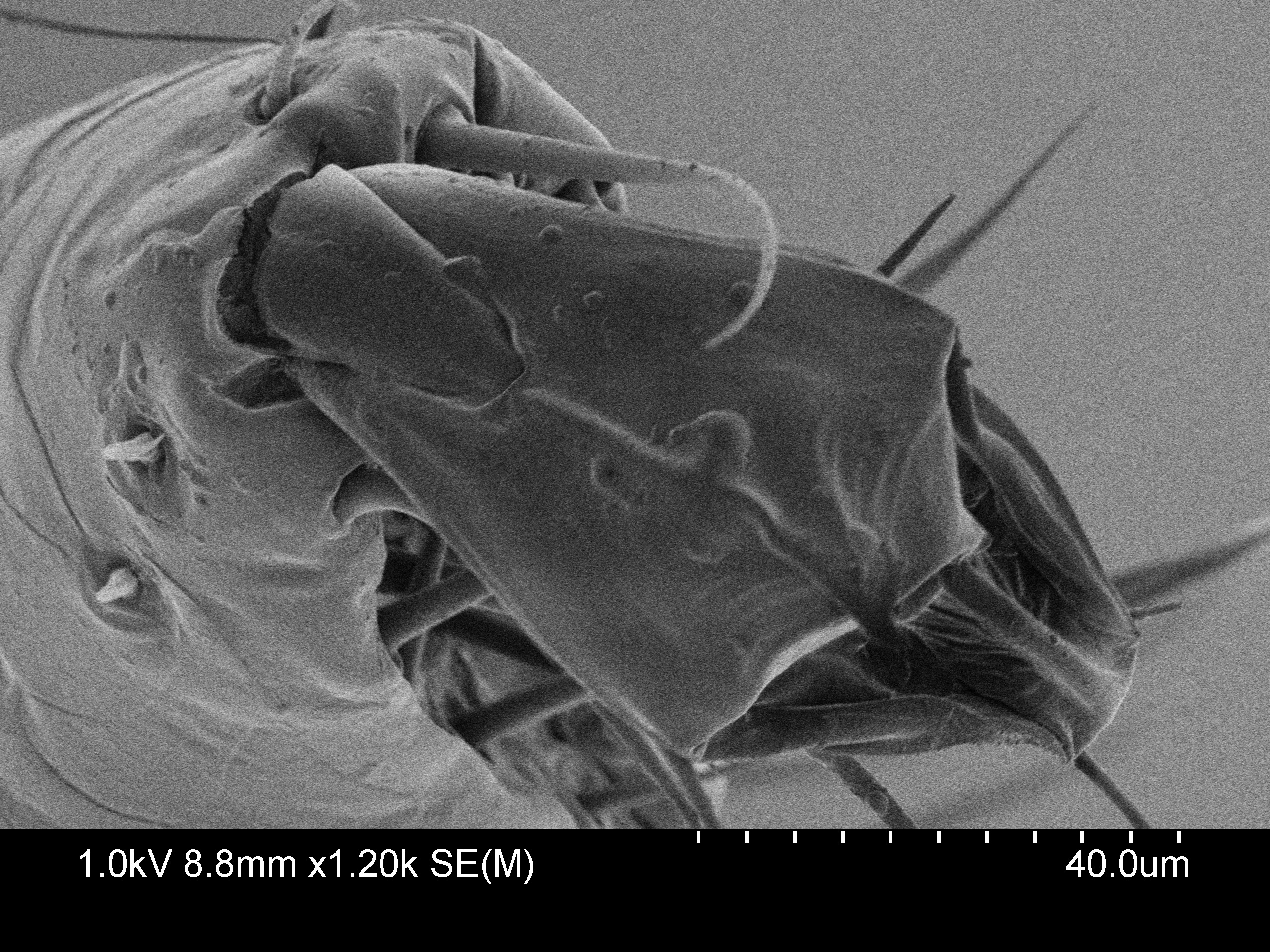
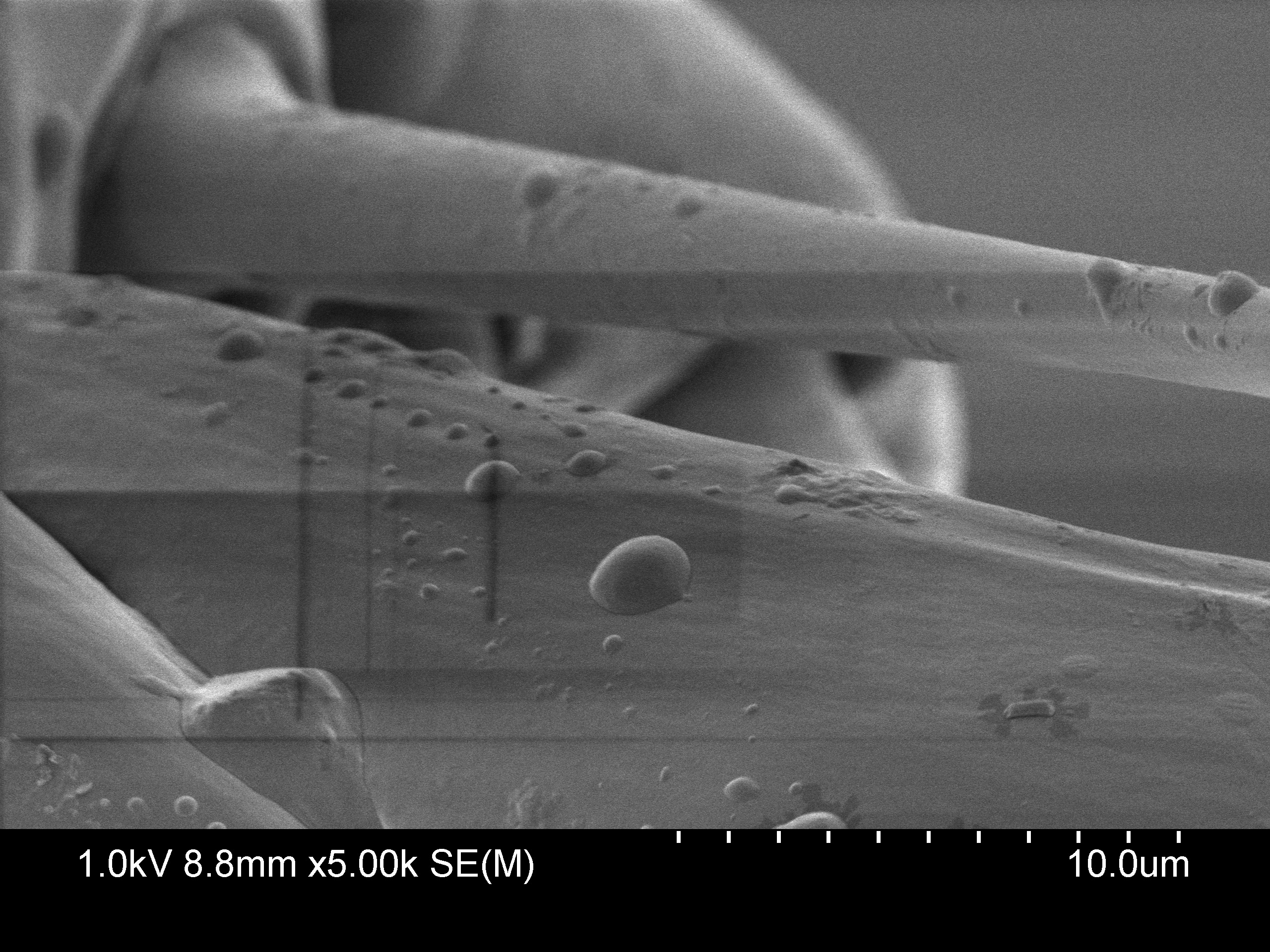
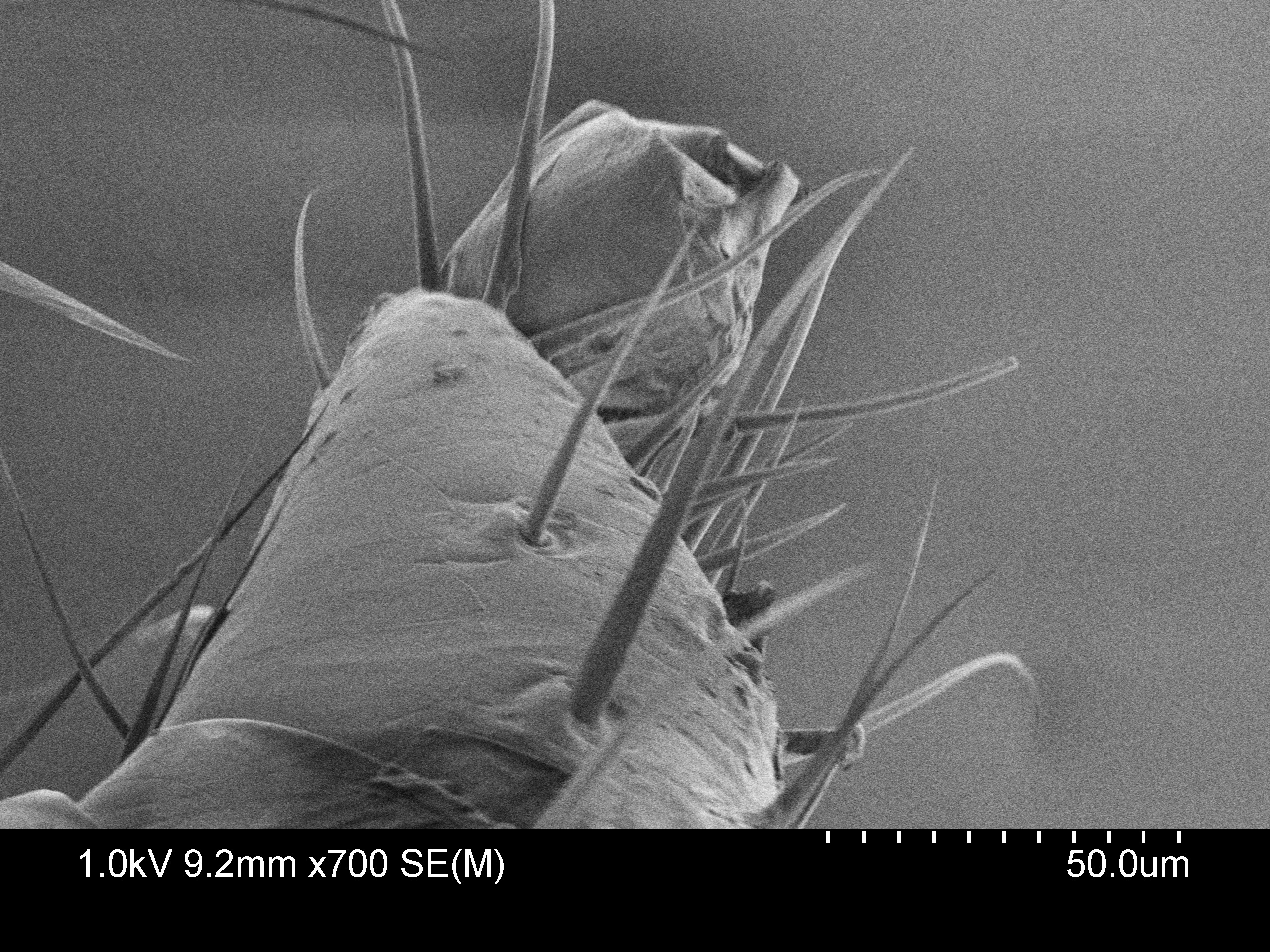
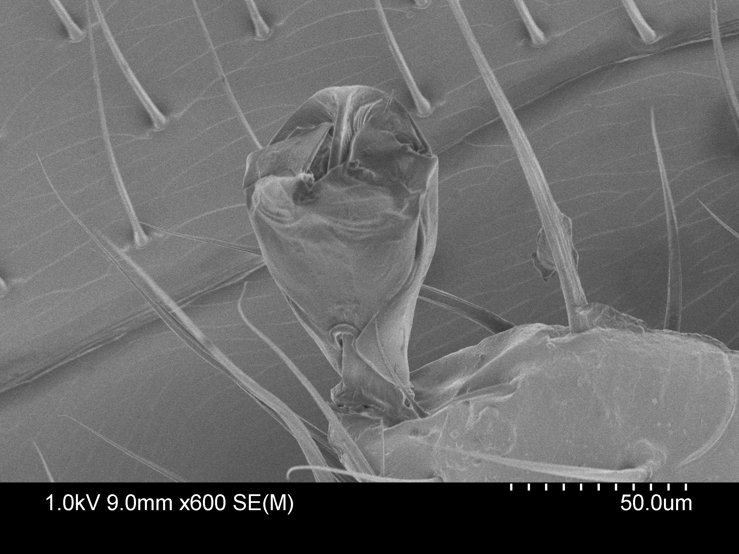
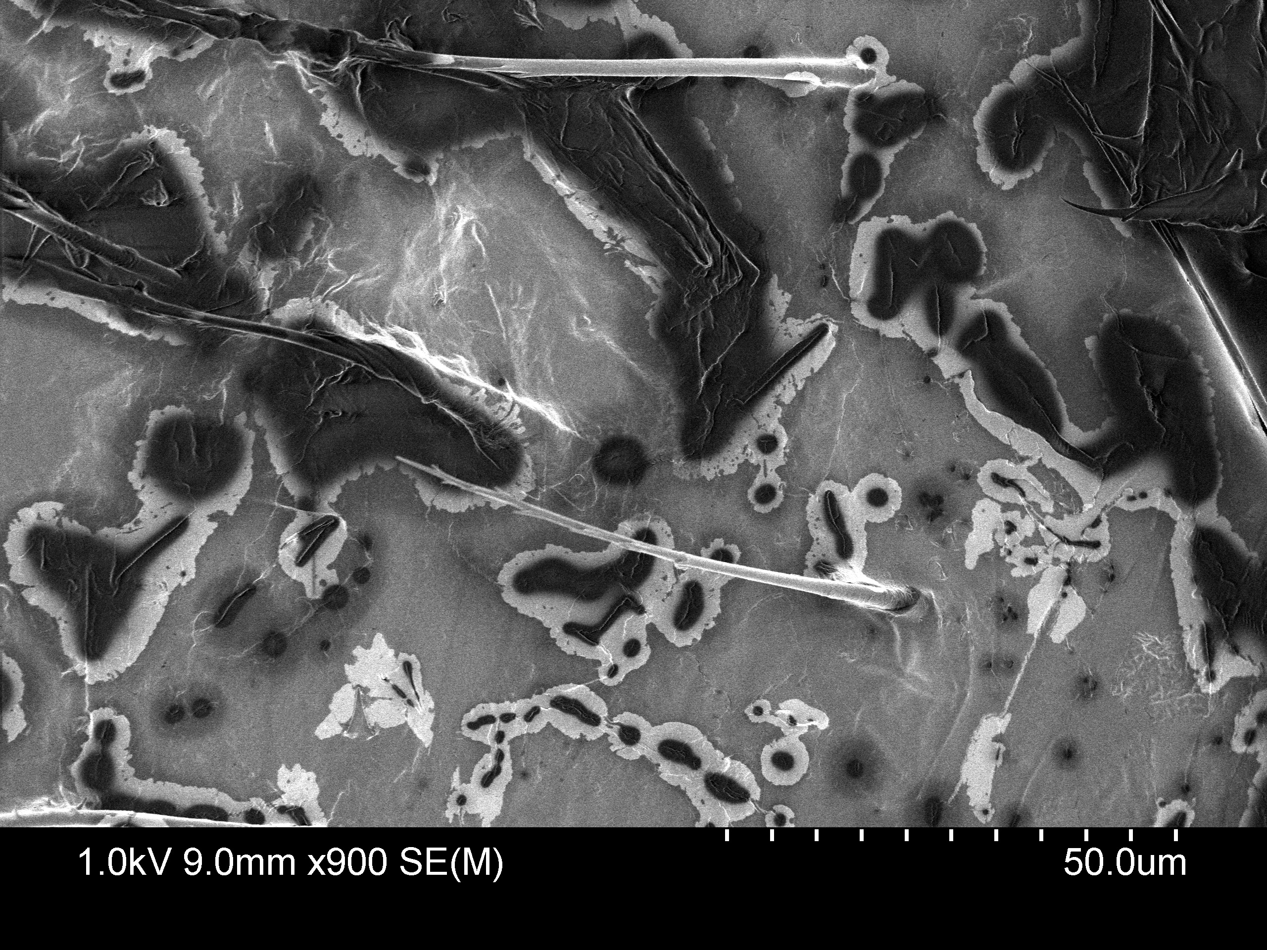
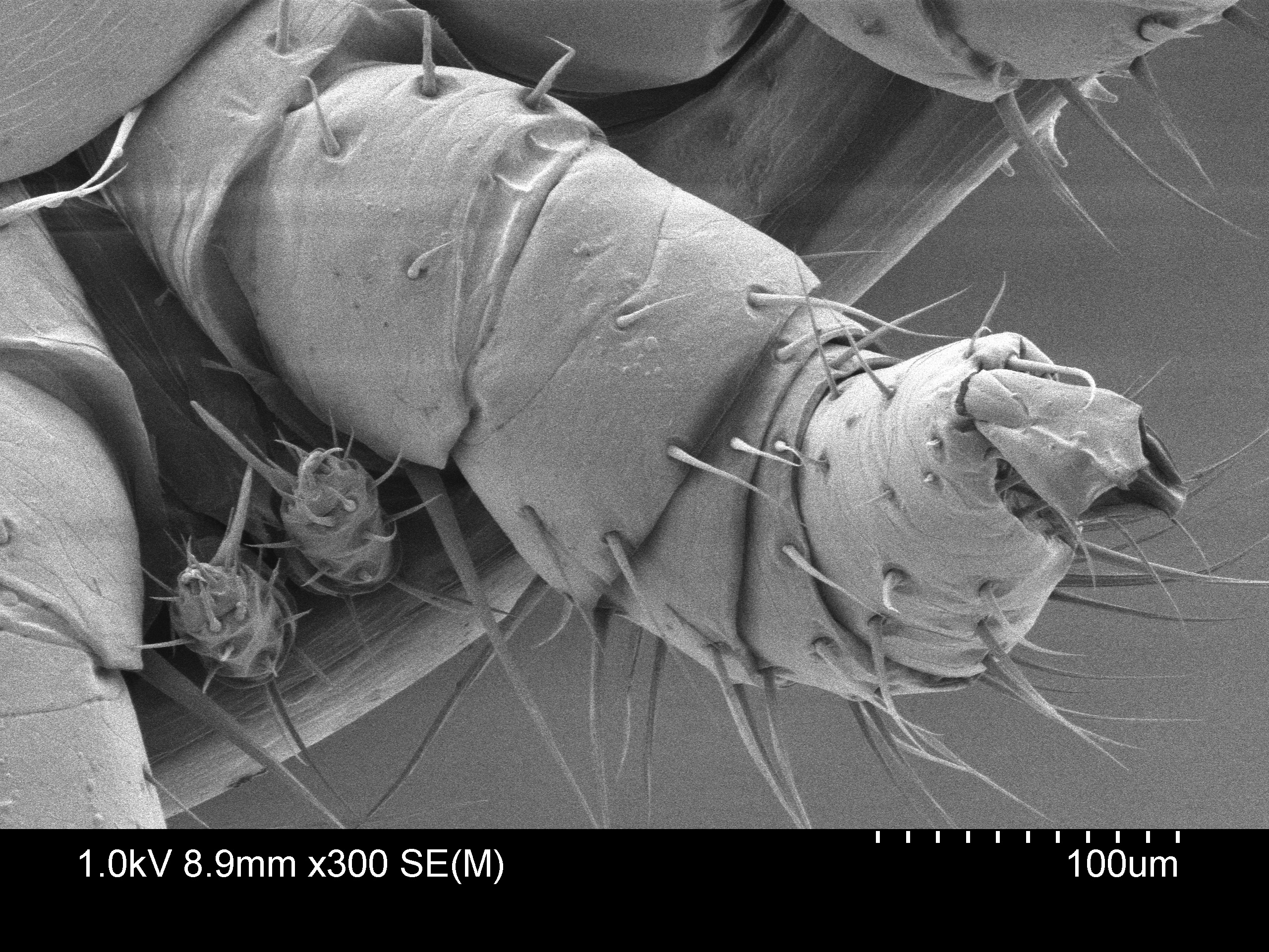
Dr. Humphrey has provided the following images she has taken using Scanning Electron Micrography of the Varroa mites that are parasitic in honey bees: ( Varroa mites (Varroa destructor and V. jacobsoni) )colonies.
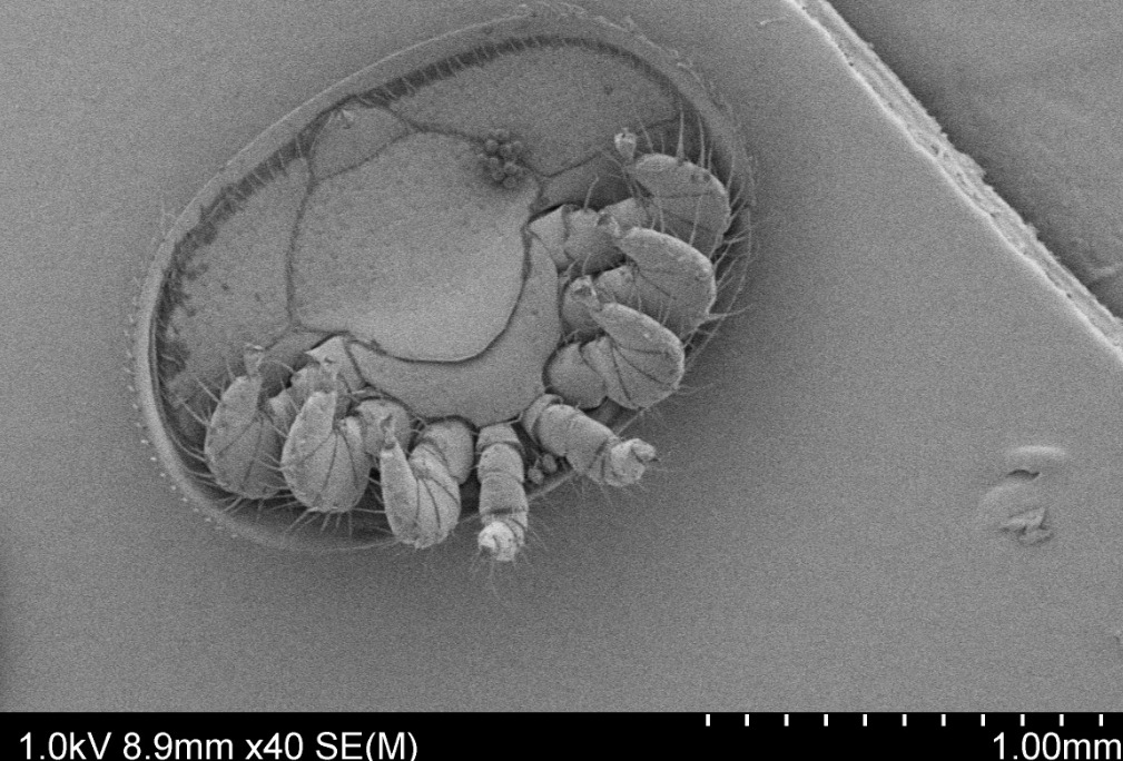
These mites are similar in name only to the mites that infect Mason bee tubes, Chaetodactylus krombeini, (Krombein’s hairy-footed pollen mite).
According to Wikipedia: ” The common name is somewhat misleading, as pollen mites consume more nectar than pollen. Both their feeding habits and their size differ significantly from Varroa destructor, the mite very harmful to the European Honey Bee. Although both are classified as mites, varroa mites are more closely related to ticks and only distantly related to Chaetodactylus. ”
So you can expect to have hairy-footed pollen mite contamination if you are not conscientious about cleaning out your tubes in the fall. See this reference on mason bee mites: